
Whether you are planning a defence medical examination, preparing for a job with strict vision standards, or simply curious whether your LASIK surgery leaves a permanent trace, here is the straight answer: yes, LASIK can be detected five years later — and far beyond that. The structural changes LASIK makes to the cornea are permanent, and an experienced eye examiner using the right instruments can identify them decades after the procedure.
What matters is how the detection happens, what specific findings give it away, and why this is clinically important rather than something to worry about. This guide from Visual Aids Centre explains the exact markers that reveal past LASIK — from corneal curvature signatures to micro-scars at the flap edge — and why being upfront about your surgical history during any future eye assessment is always the right call.
Key Takeaways
- LASIK produces permanent changes to corneal shape and thickness that remain detectable 5, 10, even 20+ years after surgery.
- Corneal topography, keratometry, and high-magnification slit-lamp examination are the primary tools used to identify past LASIK.
- The corneal flap edge leaves a faint circular signature visible under retroillumination, even after complete epithelial healing.
- Never attempt to hide past LASIK during medical examinations — disclosure protects your eligibility and ensures accurate future care.
The Short Answer — And Why It Matters
LASIK is detectable after 5 years because it is not a cosmetic overlay that fades — it is a structural reshaping of the cornea that stays with you for life. The question itself tends to come up for one of three reasons: patients preparing for defence medicals, candidates applying to roles with specific vision standards, or individuals simply curious about the permanence of the procedure.
For armed forces, civil aviation, and certain government service recruitments, examiners are specifically trained to identify post-refractive corneas. The detection methods used in medical boards are straightforward and reliable. Attempting to conceal past LASIK is not only futile but can disqualify an otherwise eligible candidate for dishonest disclosure.
What LASIK Permanently Changes in the Eye
LASIK makes three permanent alterations to the cornea that form the basis of long-term detection.
First, the central cornea becomes flatter than nature designed it. To correct myopia, the excimer laser removes stromal tissue from the central optical zone, reducing the cornea’s curvature. This new flatter profile shows up clearly on any topographic map, producing a characteristic “central flattening” pattern that does not exist in unoperated eyes. For details on the diagnostic tool used to capture these maps, see our guide on the Pentacam imaging test.
Second, the overall corneal thickness is reduced. A typical pre-LASIK cornea measures around 540 microns centrally; after surgery, the central thickness drops by 50 to 100 microns depending on the prescription corrected. Pachymetry — an ultrasound or optical measurement of corneal thickness — instantly reveals this reduction when the numbers are compared against normative data for your age and axial length.
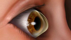
Third, the flap interface itself leaves a permanent micro-scar at the peripheral ring where the flap edge meets the underlying stroma. The flap re-adheres fully but never fuses at a molecular level, and this circular interface is visible under slit-lamp retroillumination as a faint ring around the treated zone. Even after clinical scarring becomes imperceptible to the naked eye, the structural signature remains.
How LASIK Is Detected During an Eye Exam
Corneal Topography
This is the single most reliable detection method. A topographer maps thousands of points across the corneal surface and produces a colour-coded elevation map. Post-LASIK corneas show a distinctive flat central zone surrounded by a steeper periphery — a pattern that simply does not occur naturally. Any ophthalmologist can identify this within seconds of reviewing the scan.
Keratometry
Keratometry measures the curvature of the central cornea in dioptres. Normal values sit between 42 and 46 D for most adults. Post-LASIK patients who were myopic often show readings below 40 D — an immediate red flag. However, post-LASIK keratometry can be misleading if interpreted in isolation, which is why surgeons combine it with topography and pachymetry for full context.
Slit-Lamp Examination
Under high magnification and oblique illumination, the flap edge can be seen as a thin circular line roughly 8 to 9 mm in diameter. This is particularly clear when the examiner uses retroillumination — shining light off the retina and back through the cornea. The flap ring persists indefinitely.
Anterior Segment OCT
Optical coherence tomography produces cross-sectional images of the cornea with micron-level resolution. The LASIK flap appears as a visible interface line in the anterior stroma even decades after surgery. This is the gold standard for confirming past laser surgery when other findings are ambiguous.
What a Detection Exam Looks Like at the 5-Year Mark
By the five-year mark, the flap has healed completely at the surface. Any micro-epithelial irregularities have smoothed over, and the cornea appears optically clear on casual examination. However, none of the underlying structural changes have reversed. A trained examiner looking for refractive surgery evidence will find it through:
A topography scan showing central flattening, a pachymetry reading below age-expected norms, a keratometry value inconsistent with the patient’s axial length, and a visible flap ring on slit-lamp retroillumination. These four findings in combination are essentially diagnostic. In most defence and aviation medical protocols, examiners specifically look for this cluster during recruitment screening. The fact that LASIK remains visible is also why candidates should verify whether their target role permits corrected vision — our guides on LASIK for commercial pilots and other defence roles cover the specific acceptability criteria.
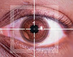
Does LASIK Affect Iris Scans and Biometric Tests?
A common concern is whether LASIK interferes with iris recognition systems used at airport immigration, corporate security, or Aadhaar verification. The short answer is no — LASIK reshapes the cornea, not the iris. The iris’s unique pattern of crypts, furrows, and pigment remains completely unchanged by laser vision correction.
However, in very rare cases, patients with significant post-LASIK dry eye or residual higher-order aberrations may experience occasional iris scan failures due to surface tear film instability scattering the infrared light used by biometric scanners. This is typically resolved by instilling a lubricating drop before scanning. Our article on LASIK and iris recognition covers this in detail.
Why You Should Always Disclose Your LASIK History
Disclosure matters for three practical reasons. First, any future cataract surgery you may eventually need — whether at 60, 70, or 80 — requires accurate keratometry readings to calculate the correct intraocular lens power. Post-LASIK corneas skew these calculations, and surgeons must use specialised formulas to avoid refractive surprises during cataract surgery. Concealing your history leads to incorrect lens selection and suboptimal vision.
Second, certain post-LASIK changes — like reduced corneal hysteresis or altered biomechanical behaviour — affect how intraocular pressure readings are interpreted. Without disclosure, your glaucoma risk assessment can be inaccurate. Third, during any future refractive enhancement, the surgeon needs complete knowledge of your original flap thickness and treatment to plan safely. Concealment in this context is directly dangerous.
Conclusion
LASIK is permanent, and its signatures in the cornea remain detectable well beyond five years — often for life. Corneal topography, pachymetry, keratometry, and slit-lamp retroillumination together provide ophthalmologists with a reliable toolkit for identifying past refractive surgery. This permanence is not a flaw — it is why LASIK works. The correction stays with you. If you are preparing for a medical examination, planning cataract surgery later in life, or want a comprehensive assessment of your post-LASIK cornea, book a consultation at Visual Aids Centre for a detailed evaluation.
Frequently Asked Questions (FAQs)
Can LASIK be detected without specialised equipment?
Not reliably. A casual eye exam with a flashlight will miss it. Detection requires a slit lamp, topographer, or anterior segment OCT — all of which are standard in any ophthalmology clinic.
Does LASIK show up on iris recognition systems?
No. LASIK reshapes the cornea, not the iris. Iris biometric systems remain fully functional after surgery in nearly all patients.
Can a doctor tell if I had LASIK 10 years ago?
Yes. The structural changes LASIK makes — central corneal flattening, reduced thickness, flap-edge ring — are permanent and detectable decades after the procedure.
Will LASIK affect my future cataract surgery?
It affects the calculations, not the feasibility. Your cataract surgeon needs to know about prior LASIK to select the right intraocular lens using post-refractive IOL formulas.
Can LASIK be hidden during a defence medical exam?
No, and attempting to do so is grounds for disqualification. Medical boards use topography and slit-lamp exams that reliably identify post-LASIK corneas. Always declare upfront.
Does LASIK leave a visible scar?
Not to the naked eye. The flap interface produces a faint ring visible only under slit-lamp retroillumination at high magnification. It does not affect vision or appearance.
👁️ MEDICALLY REVIEWED BY
Padmashree Dr. Vipin Buckshey
Optometrist & Refractive Diagnostics Specialist | AIIMS Graduate, 1977 | Padma Shri Honouree
With more than four decades of clinical experience and over 250,000 laser vision correction procedures performed at Visual Aids Centre, Dr. Vipin Buckshey routinely conducts post-refractive corneal evaluations — including detection assessments for defence medical boards and IOL calculations for patients with prior LASIK. An AIIMS alumnus, former President of the Indian Optometric Association, and official optometrist to the President of India, Dr. Buckshey ensures every diagnostic workup at the centre is clinically rigorous and honestly communicated. Learn more about our story.