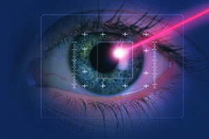

If you are asking how to detect LASIK surgery, you are probably asking for one of two reasons. Either you are a medical professional or examiner who needs to assess whether a patient’s cornea has been surgically altered — or you are a candidate for a government, defence, or professional medical examination wondering whether your procedure will be discovered. In both cases, the answer is the same: LASIK surgery is detectable with complete reliability. There is no procedure variation, no time elapsed, and no intervention that prevents a trained ophthalmologist from identifying that LASIK has been performed.
This guide from Visual Aids Centre explains exactly how LASIK is detected, which diagnostic instruments are used, what each test reveals, and why the question of concealment is not just ineffective — it is also the wrong approach for any candidate navigating a professional medical examination.
Key Takeaways
- LASIK surgery is 100% detectable by a trained ophthalmologist. No form of laser refractive surgery leaves the cornea in its pre-operative state.
- The primary detection method is slit lamp biomicroscopy — the standard clinical examination that any ophthalmologist performs. The corneal flap edge is visible as a distinct circumferential ring even years after surgery.
- Advanced instruments — Pentacam, corneal topography, wavefront aberrometry — can confirm LASIK and quantify the degree of correction applied with high precision.
- Corneal thickness reduction is a permanent, measurable, and diagnostic finding that confirms ablative corneal surgery regardless of which technique was used.
- Surface ablation procedures (PRK, ASA) are harder to detect than flap-based LASIK because they create no visible flap edge — but corneal topographic changes and thickness reduction remain detectable by experienced examiners using advanced equipment.
Is LASIK Surgery Always Detectable?
Yes — without exception. LASIK creates permanent, measurable changes to the cornea that cannot be reversed, masked, or hidden from a competent clinical examination. The two primary permanent changes are the corneal flap interface — a distinct structural plane between the flap tissue and the underlying stroma — and the reduction in overall corneal thickness resulting from the ablation performed beneath the flap.
The flap interface remains visible under slit lamp examination for life, presenting as a circumferential ring on the corneal surface. The corneal thickness reduction is quantified by Pentacam or ultrasound pachymetry with precision measured in microns. Neither finding disappears with time, and neither can be altered by any post-operative intervention short of corneal transplantation. The question “can LASIK be detected” has only one clinical answer: yes, always.
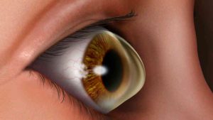
Method 1: Slit Lamp Biomicroscopy — Primary Detection
The slit lamp is the standard examination instrument in any ophthalmic clinical setting — the instrument an ophthalmologist uses in every routine eye examination. Under slit lamp illumination, the LASIK corneal flap edge is visible as a distinct circumferential line on the anterior corneal surface. This line represents the boundary between the flap tissue and the surrounding uncut corneal epithelium and stroma.
This finding is present immediately after surgery and remains indefinitely. Years after LASIK — even decades later — the flap edge remains identifiable under slit lamp examination by any trained eye specialist. The instrument required to see it is standard clinical equipment present in every ophthalmology practice. No specialised facility or advanced imaging system is needed to confirm flap-based LASIK from basic slit lamp examination alone.
Additionally, slit lamp examination reveals other LASIK indicators: subtle anterior stromal interface haze in some patients, epithelial ingrowth at the flap margin in others, and the characteristic topographic curvature changes that produce different light reflex patterns compared to an unoperated cornea. Each of these is identifiable to an experienced examiner without requiring digital quantification.
Method 2: Pentacam and Advanced Tomography
The Pentacam is a Scheimpflug-based rotating camera system that captures detailed cross-sectional images of the anterior segment and generates precise measurements of corneal thickness at every point across the corneal surface. In the context of LASIK detection, it provides two definitive findings.
First, it generates a complete pachymetric map — a colour-coded topographical representation of corneal thickness — that shows the characteristic thinning pattern produced by LASIK ablation. The central thinning relative to the peripheral cornea follows a specific ablation profile that is diagnostically distinct from natural thin cornea presentations. Second, the Scheimpflug cross-sectional image directly visualises the flap interface as a density change within the corneal stroma — confirming not just that surgery was performed but at what depth and with what ablation geometry.
Advanced diagnostic systems like Pentacam are standard equipment in specialist refractive surgery centres and are routinely used for pre-operative assessments. Our guide to the Pentacam test for LASIK explains in detail what the Pentacam measures, how the data is interpreted, and why it is considered the gold standard for pre-operative corneal assessment — as well as the most definitive post-operative detection instrument.
Method 3: Corneal Topography
Corneal topography maps the curvature of the anterior corneal surface using reflected light patterns to generate a colour-coded curvature map. A LASIK-treated cornea shows a characteristic central flattening pattern for myopic corrections — a large circular zone of reduced curvature that is absent in any naturally occurring corneal shape. For hyperopic corrections, the pattern is reversed: a steepening of the central cornea with a corresponding change in the surrounding optical zone.
These topographic signatures are not merely suggestive — they are diagnostically conclusive for any examiner experienced in reading corneal topographic maps. The ablation zone boundary is clearly demarcated as a ring of transition between the treated central zone and the untreated peripheral cornea. This boundary corresponds to the optical zone of the laser treatment and is one of the most reliable indicators of prior laser refractive surgery across all procedure types.
Our overview of the corneal topography test explains how the mapping works, what different curvature patterns indicate, and how examiners distinguish normal corneal variations from post-surgical topographic changes.
Method 4: Wavefront Evaluation
Wavefront aberrometry measures the optical quality of the eye’s complete optical system — identifying deviations from perfect optical performance called higher-order aberrations. LASIK surgery changes the corneal optical profile in ways that produce specific wavefront signatures, particularly in the transition zones between the ablated optical zone and the untreated corneal periphery.
Post-LASIK wavefront maps typically show increased spherical aberration — the most characteristic wavefront signature of laser corneal ablation. The pattern and magnitude of spherical aberration change is proportional to the correction applied and the ablation profile used. Wavefront-guided LASIK reduces this signature but does not eliminate it. A trained wavefront analyst can identify prior laser surgery from the aberrometry map even without access to the patient’s surgical history.
Wavefront evaluation also identifies higher-order aberrations that contribute to patient-reported visual quality concerns after LASIK — halos, glare, reduced contrast. Our resource on wavefront-guided LASIK explains how the wavefront data is used both for detecting prior surgery and for optimising future corrections where enhancement procedures are considered.
Method 5: Dry Eye Examination
LASIK surgery disrupts the corneal sensory nerve network responsible for triggering tear secretion. The resulting nerve regeneration follows a characteristic timeline and pattern. Dry eye testing in a patient with prior LASIK may reveal specific patterns of nerve-related tear film instability that are diagnostically informative to an experienced clinician.
The standard dry eye diagnostic battery includes tear break-up time (TBUT) assessment, Schirmer testing of aqueous tear production, lipid layer evaluation, and meibography. Meibography — imaging the meibomian glands using infrared photography — is increasingly incorporated into comprehensive LASIK screening because meibomian gland dysfunction is both a LASIK risk factor and a post-LASIK complication contributor. Collectively, these tests support the detection picture assembled from topography and biomicroscopy. For patients managing ongoing dry eye after refractive surgery, our resource on the Schirmer test explains what this quantitative tear assessment involves and how results guide ongoing management.
Method 6: Contrast Sensitivity and Pupil Evaluation
Contrast Sensitivity Testing
Standard visual acuity charts measure only high-contrast resolution — whether you can distinguish black letters on a white background. Contrast sensitivity testing evaluates the eye’s ability to detect objects against progressively similar-toned backgrounds, measuring functional vision quality in conditions closer to real-world environments. LASIK patients — particularly those with significant ablations or large scotopic pupil sizes — may show characteristic contrast sensitivity reductions in the mid-frequency range that are diagnostically informative in a post-operative context.
Pupil Evaluation
Pupil diameter under low-light (scotopic) conditions is a relevant detection parameter for several reasons. Patients with larger scotopic pupils relative to their laser ablation zone are known to experience more pronounced halos and glare — a pattern that can be identified in the post-operative setting. Infrared pupilometry provides precise pupil measurement under standardised conditions. The relationship between pupil size, ablation zone diameter, and optical side effects is a clinically informative combination that contributes to the comprehensive detection assessment.
How Detectable Is PRK Compared to LASIK?
Photorefractive Keratectomy (PRK) and its derivative advanced surface ablation (ASA) are harder to detect than flap-based LASIK because they create no corneal flap and therefore no visible flap edge under slit lamp examination. However, “harder to detect” is not the same as “undetectable.” The corneal topographic changes — the central flattening or steepening produced by the ablation — are present in PRK-treated corneas in exactly the same way as in LASIK-treated corneas. The corneal thickness reduction is also present and quantifiable by pachymetry.
What changes with surface ablation is the slit lamp detection method — the examiner looks for subtle anterior surface changes and characteristic topographic patterns rather than a visible flap edge. For an experienced refractive surgery examiner using Pentacam or topography, prior PRK remains identifiable. It requires more expertise and better equipment than LASIK detection — but it is not invisible. Our resource on keratoconus treatment gives helpful context on how corneal tomography distinguishes ectatic disease from post-surgical corneal shape changes — the same diagnostic framework applied in LASIK detection for candidacy screening.
Why Disclosure Is Always the Right Approach
For candidates appearing for government, defence, or professional medical examinations: attempting to conceal prior LASIK surgery is not only ineffective but carries independent consequences that far exceed the risk of honest disclosure. The detection methods described in this article are standard clinical practice. An ophthalmologist conducting a medical examination for a government service or defence recruitment board will apply slit lamp examination as a minimum — and the flap edge, if present, will be seen.
Concealment, when discovered, is treated as misrepresentation of medical history — a disqualifying act independent of the vision outcome or procedure type. Many services that previously restricted LASIK have updated their policies to accommodate post-LASIK candidates who meet the vision standard and disclose their surgical history honestly. The conversation about whether your post-LASIK vision qualifies you is a conversation worth having. The conversation about whether your concealment was caught is not one you want to have. Our guide on Contoura and topography-guided LASIK covers how these procedure variations differ in their topographic signatures — relevant for candidates or examiners assessing which type of refractive surgery was performed.
Conclusion
LASIK surgery is detectable by any trained ophthalmologist using equipment that is standard in every eye clinic. The slit lamp reveals the flap edge. Pentacam quantifies the corneal thickness reduction and visualises the ablation pattern. Topography maps the characteristic curvature changes. Wavefront aberrometry identifies the post-surgical optical signature. No elapsed time, no subsequent procedure, and no technique variation renders LASIK undetectable to a competent examiner.
For anyone considering LASIK and navigating questions about professional or government medical examinations — the right approach is honest disclosure, not concealment. Book a consultation at Visual Aids Centre to understand what your post-operative corneal profile reveals clinically and how to navigate your specific medical examination context with accurate documentation.
Frequently Asked Questions (FAQs)
Can LASIK surgery be detected years after the procedure?
Yes, permanently. The corneal flap edge remains visible under slit lamp examination for life, and the corneal thickness reduction is measurable by pachymetry indefinitely. No amount of elapsed time reduces the detectability of flap-based LASIK.
What instrument is used to detect LASIK?
The primary instrument is the slit lamp biomicroscope — standard equipment in every ophthalmology clinic. Advanced instruments including Pentacam, corneal topographers, and wavefront aberrometers provide additional quantitative confirmation. A basic slit lamp examination is sufficient to identify flap-based LASIK in almost all cases.
Can PRK or surface ablation procedures be detected?
Yes, though detection requires more sophisticated equipment than LASIK because there is no flap edge to identify. Corneal topographic changes and thickness reduction remain present and quantifiable by Pentacam and topography. An experienced examiner using advanced imaging can identify prior surface ablation with high reliability.
Will LASIK be detected in a government or defence medical examination?
Yes. Medical examinations for government, defence, and security services are conducted by ophthalmologists who apply standard slit lamp examination as a minimum. The LASIK flap edge is consistently identified by trained examiners. Attempting to conceal prior LASIK is ineffective and constitutes misrepresentation of medical history.
Does Pentacam detect LASIK more accurately than slit lamp?
Pentacam provides more quantitative detail — precise corneal thickness maps, ablation depth estimation, and Scheimpflug cross-sectional imaging of the flap interface. The slit lamp provides direct visual confirmation of the flap edge. Both are reliable detectors; Pentacam adds quantification while slit lamp provides the definitive clinical observation.
Can wavefront testing detect that LASIK was performed?
Yes. Post-LASIK corneas show characteristic wavefront signatures — particularly increased spherical aberration — that are diagnostically informative to an experienced analyst. Wavefront aberrometry contributes to the comprehensive post-LASIK detection picture alongside topography and slit lamp findings.
👁️ MEDICALLY REVIEWED BY
Padmashree Dr. Vipin Buckshey
BS Ophthalmology | AIIMS Graduate, 1977 | Padma Shri Honouree | Corneal Diagnostics and Refractive Surgery Detection Specialist, Visual Aids Centre
The clinical examination skills that allow an experienced ophthalmologist to detect prior LASIK surgery in seconds under a slit lamp are built over years of examining hundreds of post-operative corneas. Dr. Vipin Buckshey’s four decades of refractive surgery practice at Visual Aids Centre — spanning the full history of laser refractive surgery from its earliest clinical adoption — have given him direct experience with every generation of LASIK technique and the clinical signatures each leaves behind. His diagnostic perspective informs this article’s explanation of what each instrument reveals and why certain procedure variations are more or less readily identified. This is not theoretical knowledge — it is built from examining post-operative corneas as a practising refractive surgeon whose own patients he follows longitudinally. An AIIMS alumnus, Padma Shri honouree, and former President of the Indian Optometric Association. Learn more about our clinical standards at our story.