
One of the most reassuring facts about LASIK is that results are typically clear and stable within days. One of the most underappreciated facts is that the cornea is still healing for months after that clarity arrives. These two things are not contradictory—they reflect the layered nature of corneal repair, where functional recovery happens fast but biological healing unfolds gradually. Understanding this distinction matters for every patient, because the decisions you make during the extended healing window directly shape how well your corneal tissue ultimately bonds, stabilises, and protects your long-term visual outcome.
This guide explains exactly how the cornea heals after LASIK, what the flap and the underlying stroma do during each phase, which complications can disrupt the process, and how to give your cornea the best possible environment to recover fully.
Key Takeaways
- The corneal epithelium seals the flap edge within 24–48 hours, but full stromal bonding takes up to three months.
- Corneal nerves—disrupted by the flap incision—regenerate gradually over 6–12 months, which is why dry eye symptoms persist beyond early recovery.
- Flap complications such as epithelial ingrowth, diffuse lamellar keratitis (DLK), and striae are rare but recognised; early detection and treatment produce good outcomes.
- Flapless procedures like SMILE Pro preserve more corneal nerve fibres and structural tissue, resulting in faster biological healing.
- Simple protective habits—avoiding eye rubbing, wearing sunglasses, using prescribed drops—directly support the corneal repair process during the full healing window.
What Happens to the Cornea During LASIK
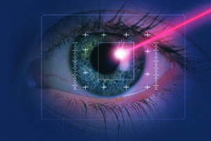
To understand how the cornea heals, it helps to understand precisely what LASIK does to it. A microkeratome or femtosecond laser creates a thin hinged disc of corneal tissue—the flap—by cutting through the epithelium and into the superficial stroma while leaving one edge attached. The flap is then folded back to expose the deeper stromal bed, where an excimer laser removes a calculated amount of tissue to reshape the cornea’s optical profile. The flap is then repositioned and adheres to the stromal bed without sutures.
The flap’s physical characteristics—its thickness and diameter—directly influence how healing proceeds. Our dedicated articles on LASIK flap diameter explain how surgeons determine these parameters and why precision here matters for both the immediate procedure and long-term corneal integrity.
The Corneal Healing Timeline: Phase by Phase
First 24–48 Hours: Epithelial Sealing
The most rapid phase of corneal healing occurs in the first day or two. The corneal epithelium—the outermost layer of cells that covers the entire ocular surface—migrates across the exposed flap edge and seals it shut. This happens with remarkable speed because epithelial cells are among the fastest-regenerating cells in the human body. By the end of day two, the flap is physically sealed at its margins, and the initial risk of mechanical disturbance is substantially reduced. Vision often improves dramatically during this window, which surprises many patients—clarity returning while healing is still in its earliest phase.
Days 3–14: Stromal Adhesion Strengthens
With the epithelial seal in place, the deeper stromal tissue begins establishing biochemical adhesion between the flap’s undersurface and the ablated stromal bed. This is not a simple gluing process—it involves the deposition of fibronectin and other adhesion proteins, along with gradual re-integration of keratocytes (the cells responsible for maintaining corneal structure) at the wound margin. The flap is stable during normal daily activity at this point, but rubbing, direct pressure, or trauma can still displace it. This is the phase where post-operative inflammation is most likely to be clinically significant—and why anti-inflammatory eye drops are prescribed for this window.
Weeks 3–12: Ongoing Remodelling
During this longer phase, the stromal wound continues consolidating and the cornea undergoes gradual structural remodelling as it adapts to its new shape. Limbal stem cells—located at the corneal periphery—play a central role in this longer-term regenerative process. Our article on the role of limbal stem cells in post-LASIK corneal healing explains the specific mechanisms by which these cells contribute to stromal repair. By the end of three months, the flap is considered fully integrated into the corneal structure—though it will never be anatomically identical to an uncut cornea.
Nerve Regeneration: The Invisible Recovery
The healing process most patients don’t see—and the one that has the most impact on long-term eye comfort—is corneal nerve regeneration. The flap incision severs the subbasal nerve plexus, the dense network of sensory fibres just beneath the corneal epithelium that govern both sensation and, critically, the reflex signals that stimulate tear production. When these nerves are cut, blink-triggered tear secretion is temporarily reduced even while the eyes remain open—which is the underlying cause of the dry eye symptoms that so many LASIK patients experience in the months following surgery.
Nerve regeneration is gradual, typically taking 6–12 months for meaningful restoration and up to two years for complete recovery in some patients. Our article on symptoms of nerve disruption after LASIK explains what patients typically notice during this period and how to distinguish normal nerve regeneration symptoms from signs that warrant clinical attention.
How LASIK Affects Corneal Biomechanics
Beyond cellular healing, LASIK changes the mechanical behaviour of the cornea permanently. Removing stromal tissue reduces the cornea’s tensile strength, and creating a flap alters its structural load distribution. For the vast majority of patients whose corneas met pre-operative thickness criteria, these changes fall well within safe parameters and have no clinically meaningful effect. For a small group—particularly those with borderline corneal thickness or subclinical irregularities—the biomechanical change is more significant and can, in rare cases, progress to post-LASIK ectasia.
Our detailed article on how LASIK surgery affects corneal biomechanics covers the physical science behind this in full—including what measurements surgeons take pre-operatively to assess biomechanical risk, and how much tissue removal is considered structurally safe.
Flap Complications and How They Are Managed
LASIK flap complications are uncommon in experienced hands, but knowing what they are—and what the treatment looks like—removes unnecessary anxiety if one of them is detected at a post-operative review.
Epithelial Ingrowth
This occurs when epithelial cells migrate underneath the flap rather than staying at the surface. Small, peripheral ingrowths are often monitored rather than treated. Larger ingrowths that approach the visual axis and affect clarity are addressed by lifting the flap and mechanically removing the migrated cells.
Diffuse Lamellar Keratitis (DLK)
Sometimes called “sands of the Sahara,” DLK is an inflammatory reaction that develops in the interface between the flap and the stromal bed—typically in the first days after surgery. It is graded by severity; early, mild DLK usually resolves with intensified steroid drops. More advanced cases may require the flap to be lifted and the interface irrigated. Our article on diffuse lamellar keratitis after LASIK covers the presentation, grading system, and treatment approach comprehensively.
Flap Striae and Irregular Astigmatism
Striae are micro-wrinkles or folds in the flap, most often detected at the first post-operative day. Clinically insignificant striae require no intervention. Those that affect visual quality can often be resolved by floating and smoothing the flap within the first few days while the stromal bed is still amenable to repositioning. Patients who notice persistent hazing, glare, or distortion in the weeks after LASIK—beyond what dry eye typically causes—may be experiencing irregular astigmatism after LASIK, which has its own specific diagnostic and treatment pathway.
Post-LASIK Ectasia
The most serious long-term corneal complication is ectasia—progressive forward bulging of the cornea caused by insufficient structural strength following tissue removal. It is rare when proper pre-operative screening is performed, but it can occur and requires prompt intervention. Corneal cross-linking (CXL) is the primary treatment, halting progression by stiffening the remaining stromal collagen. Our article on cross-linking for post-LASIK ectasia explains how this works and when it is indicated.
Protecting the Healing Cornea
The cornea heals best when it is given a stable, low-stress environment. The instructions that surgeons provide are not formalities—they directly support the biological processes described above.
- Do not rub your eyes. Mechanical pressure on the flap during the healing window is the leading preventable cause of flap displacement and can disrupt epithelial sealing in the first days.
- Wear protective shields while sleeping for the first week. Involuntary nocturnal pressure on closed eyelids is the most common mechanism of accidental post-operative flap disturbance.
- Complete your antibiotic and anti-inflammatory drops. These address the two most significant biological threats to flap healing in the early window—infection and excessive inflammation.
- Protect the cornea from UV radiation. Ultraviolet light can slow epithelial regeneration and increase photochemical stress on healing stromal tissue. Wearing appropriate sunglasses every time you go outdoors is not optional during recovery.
- Attend all follow-up appointments. Flap complications and early ectasia are identified at post-operative reviews—often before they become symptomatic. The earlier they are caught, the simpler the management.
Does Flapless Surgery Heal Differently?
Yes—and in meaningful ways. Procedures like SMILE Pro do not create a flap at all; instead, a lenticule is extracted through a small keyhole incision. This preserves the anterior stromal lamellae and the superficial nerve plexus to a far greater degree than any flap-based technique. The result is a fundamentally different healing profile: fewer disrupted corneal nerves means less post-operative dryness, faster nerve regeneration, and a cornea that retains more of its native biomechanical properties.
Conclusion
Yes—the cornea heals after LASIK, and it does so with impressive efficiency. The epithelium seals within 48 hours, the flap achieves solid stromal adhesion within weeks, and the remodelling process completes over three months. Corneal nerve regeneration takes longer still, which is why dry eye symptoms can outlast the visible healing window. Understanding the layered nature of corneal repair helps make sense of both the speed of functional recovery and the reason certain precautions remain important well past the first week.
The rare complications associated with LASIK flaps are manageable when detected early—which is exactly why post-operative follow-up is as important as the surgery itself. At Visual Aids Centre, our post-operative protocol is designed around the corneal healing timeline described in this article, ensuring that every patient is monitored at the right intervals to catch any deviation early. If you’re considering LASIK and want to understand how your individual corneal profile affects your candidacy and recovery outlook, book a consultation with our team.
Frequently Asked Questions (FAQs)
How long does the corneal flap take to fully heal after LASIK?
The flap edge is sealed by the epithelium within 24–48 hours. Stromal adhesion strengthens progressively over the following weeks, with full integration typically considered complete around the three-month mark. The flap never becomes anatomically identical to uncut tissue, but it is firmly and permanently bonded to the stromal bed.
Can the LASIK flap ever fall off or detach permanently?
A complete flap detachment is exceptionally rare after the first week. Partial displacement from direct blunt trauma can occur during the early healing window, which is why eye rubbing and contact sports are restricted. After three months, the flap is integrated sufficiently that accidental displacement is extremely unlikely.
Why do I still have dry eyes months after LASIK?
Dryness persisting beyond the first few weeks is driven by corneal nerve regeneration, which takes 6–12 months on average. The subbasal nerve plexus—severed during flap creation—governs the reflex tear secretion triggered by each blink. As nerves regenerate, tear production normalises.
Can LASIK cause corneal scarring?
Corneal scarring after LASIK is rare in straightforward cases. It is more associated with surface ablation procedures like PRK, or with complicated healing in the context of infection or severe inflammation.
What is epithelial ingrowth and is it serious?
Epithelial ingrowth occurs when surface cells migrate beneath the flap after surgery. Small, peripheral cases are typically monitored rather than treated. Cases that encroach on the visual axis or cause flap irregularity are treated by lifting the flap and clearing the cells. When detected early, outcomes are excellent.
Does flapless LASIK heal faster than standard LASIK?
In terms of nerve regeneration and dry eye resolution, yes—flapless procedures like SMILE Pro spare more of the subbasal nerve network, resulting in faster tear function recovery. The absence of a flap also removes the risk of flap-related complications entirely.
👁️ MEDICALLY REVIEWED BY
Padmashree Dr. Vipin Buckshey
Optometrist & Post-Operative Care Specialist | AIIMS Graduate, 1977 | Padma Shri Honouree
With more than four decades of clinical experience and over 250,000 laser vision correction procedures performed at Visual Aids Centre, Dr. Vipin Buckshey has overseen the post-operative corneal healing protocols for LASIK, SMILE Pro, Contoura Vision, and Trans-PRK across thousands of procedures—including the clinical management of flap complications, ectasia, and corneal nerve regeneration in complex cases. An AIIMS alumnus, former President of the Indian Optometric Association, and official optometrist to the President of India, Dr. Buckshey personally reviews all clinical content at Visual Aids Centre to ensure it reflects both current evidence and the realities of real patient experience. Learn more about the centre’s clinical philosophy at our story.