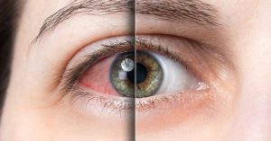
If you noticed a bright red patch on the white of your eye after LASIK, the first instinct is to worry. It looks dramatic — dark red, sometimes covering a large portion of the visible sclera — and nothing in your pre-surgery preparation quite prepares you for how it looks in the mirror the morning after. The good news is that what most patients call a “blood clot” is almost always a subconjunctival haemorrhage: a ruptured surface blood vessel under the clear conjunctival membrane. It is not a clot in the cardiovascular sense. It cannot travel anywhere. And in the overwhelming majority of cases, it needs no treatment at all.
That said, not all post-LASIK redness is the same, and understanding what you are looking at — and what would actually require urgent attention — is genuinely useful for anyone going through LASIK recovery. This article covers the clinical facts, the risk factors that make haemorrhage more likely, how long it takes to resolve, and the specific symptoms that should prompt a same-day call to your surgeon.
💡 Quick Highlights
- Post-LASIK “blood clots” are almost always subconjunctival haemorrhages — ruptured surface vessels, not vascular clots. They look alarming but are typically harmless.
- The suction ring used during flap creation places temporary pressure on the eye, which is the most common cause of vessel rupture during LASIK.
- Patients on blood-thinning medications (aspirin, ibuprofen, clopidogrel) have higher risk — always disclose these to your surgeon before surgery.
- A subconjunctival haemorrhage does not affect vision and resolves on its own in one to three weeks without treatment.
- Severe eye pain, vision loss, or haemorrhage that spreads significantly after the first day are symptoms that need urgent assessment — not watchful waiting.
In This Article
What a “Blood Clot” After LASIK Actually Is
It looks like a great deal of blood because the contrast against the white sclera is stark. The actual volume is tiny — fractions of a millilitre. The conjunctival vessels are fragile and superficial. A subconjunctival haemorrhage cannot enter the eye’s interior, cannot affect the retina, the cornea, or the visual pathway, and does not indicate bleeding elsewhere. For detailed information on this specific presentation after LASIK, our article on subconjunctival haemorrhage after LASIK covers the clinical picture in full.
This is distinct from intraocular bleeding — haemorrhage within the eye itself — which is genuinely rare after LASIK and would present with significant vision changes or severe pain, not just a red patch visible from the outside. For a broader look at LASIK’s relationship to bleeding risk, see our resource on whether LASIK causes bleeding and what the evidence actually says.
What Causes It — Surgery and Patient Factors
The Suction Ring During Flap Creation
LASIK uses a suction ring to stabilise the eye and create the vacuum needed for the femtosecond laser to create the corneal flap precisely. This ring applies significant pressure to the ocular surface — transiently but reliably raising intraocular pressure to levels that stress the delicate conjunctival vessels. For most patients these vessels hold. For some, particularly those with naturally fragile surface vasculature, one or more ruptures.
The haemorrhage typically appears in the area of the conjunctiva closest to where the suction ring contacted the eye. It is a direct mechanical consequence of the procedure — not a sign of surgical error or poor technique. SMILE Pro’s flapless design avoids the suction ring pressure of LASIK’s flap creation step, which is one reason subconjunctival haemorrhage is marginally less common with flapless procedures.
Patient-Side Risk Factors
Some patients are substantially more prone to this than others. The most clinically significant patient-related factors are:
- Blood-thinning medications: Aspirin and NSAIDs like ibuprofen reduce platelet aggregation, which directly impairs the clotting response that would normally limit vessel bleed. Prescription anticoagulants (warfarin, clopidogrel, newer anticoagulants) carry even higher risk. Most surgeons ask patients to stop aspirin and NSAIDs at least one week before surgery where medically safe to do so — always disclose all medications at your pre-operative consultation.
- Uncontrolled hypertension: Elevated blood pressure places more stress on surface vessel walls and increases rupture risk. Blood pressure should be under control in the period leading up to surgery.
- Diabetes: Diabetic microangiopathy causes fragility in small vessel walls throughout the body, including the conjunctival vessels.
- Vigorous eye rubbing before the procedure: Some patients, nervous before surgery, rub or press on the eye — this can rupture surface vessels even before the suction ring is applied.
- Valsalva events during surgery: Breath-holding, coughing, or straining on the operating table temporarily raises systemic venous pressure and can rupture surface vessels.
Warning Signs That Need Same-Day Attention
A subconjunctival haemorrhage on its own — a red patch, no pain, normal vision — does not require urgent assessment beyond your scheduled post-operative review. The appearance alone is not an emergency. What changes the picture:
⚠️ Contact Visual Aids Centre the same day if you notice:
- Significant or worsening eye pain — not the normal dryness-type discomfort, but persistent aching or sharp pain
- Any change in visual acuity — blurring, distortion, or a reduction in the clarity you had at the one-day post-op check
- The haemorrhage expanding noticeably beyond the first 24 hours, particularly if it spreads toward the cornea
- Haemorrhage alongside symptoms of raised intraocular pressure: eye feels “tight” or hard to the touch, rainbow halos around lights, nausea
- Discharge alongside redness — this combination suggests infection rather than simple vessel rupture
The distinction between the common, benign presentation and the less common concerning one is worth holding clearly in mind. Bloodshot eyes in general during early recovery are discussed in our resource on bloodshot eyes after LASIK surgery, which covers the full range from mild conjunctival redness to presentations worth acting on.
How Long It Takes to Resolve
The haemorrhage resolves as the pooled blood is gradually absorbed by surrounding tissue. The typical progression looks like this:
The patch often looks its worst — maximum colour intensity, sometimes appearing to have slightly spread as blood redistributes. This is normal and does not indicate ongoing bleeding.
Colour begins shifting from bright red toward orange or yellow at the edges as haemoglobin breaks down. The overall size usually stabilises or begins reducing.
Gradual fading. Most subconjunctival haemorrhages clear completely within two to three weeks. Larger ones, or those in patients with impaired clotting, may take a few days longer.
For a specific look at how long the visible red spots and discolouration typically take to clear after LASIK, our article on how long it takes for red spots to go away after LASIK maps the healing timeline in more detail.
Reducing the Risk Before and After Surgery
You cannot eliminate the risk of subconjunctival haemorrhage after LASIK entirely — it is partly a consequence of the suction ring mechanics. But you can reduce it:
Before surgery: Stop aspirin and non-prescription NSAIDs at least seven days before the procedure if your prescribing physician confirms this is safe. Tell your surgeon about all medications at your pre-operative consultation, including supplements like vitamin E, fish oil, and ginkgo, which have mild anticoagulant properties. Ensure blood pressure is controlled if you have hypertension.
On the day: Avoid rubbing your eyes, straining, or anything that raises venous pressure significantly before the procedure. Stay calm — easier said than done, but the Valsalva effect from tensing up on the operating table is a real contributing factor.
After surgery: Follow the prescribed drop schedule precisely. Antibiotic and anti-inflammatory drops after LASIK are not primarily for the haemorrhage — they are for infection prevention and healing support — but they are part of the comprehensive care that supports overall post-operative recovery. Our resource on using eye drops after LASIK covers the full post-operative drop schedule and what each drop does.
Frequently Asked Questions
Is a blood clot after LASIK dangerous?
In almost all cases, no. What patients call a “blood clot” is a subconjunctival haemorrhage — a surface vessel rupture. It cannot affect vision, cannot enter the eye’s interior, and resolves on its own without treatment. The situations that are genuinely concerning are those accompanied by pain, vision change, or signs of raised intraocular pressure — none of which are typical of a simple subconjunctival haemorrhage.
How long does a blood clot take to go away after LASIK?
Most subconjunctival haemorrhages clear completely within one to three weeks. The patch typically looks worst in days one to three, then gradually changes colour from red to orange-yellow as the blood breaks down, before fading entirely. Larger patches or those in patients with slow clotting may take slightly longer.
Will the haemorrhage affect my LASIK results?
No. A subconjunctival haemorrhage involves the conjunctiva — the membrane covering the white of the eye. LASIK reshapes the cornea, which is entirely separate. The haemorrhage does not affect the surgical correction, the corneal healing process, or your final visual outcome.
Should I stop taking aspirin before LASIK?
Generally yes — most surgeons recommend stopping aspirin and other NSAIDs at least seven days before surgery to reduce bleeding risk. This must be done with your prescribing physician’s agreement, particularly if aspirin is part of a cardiovascular prevention regimen. Never stop a prescribed medication without medical clearance.
Is a subconjunctival haemorrhage the same as a blood clot?
They are different things. A subconjunctival haemorrhage is blood pooling under a surface membrane from a ruptured vessel — it stays localised, cannot move, and carries no systemic risk. A blood clot in the vascular sense (such as a deep vein thrombosis) is a formed clot within a blood vessel. The two terms are used interchangeably by patients but describe entirely different clinical events. LASIK does not cause vascular blood clots.
Can I use eye drops if I have a subconjunctival haemorrhage after LASIK?
Yes — continue your prescribed antibiotic and anti-inflammatory drops on schedule regardless of the haemorrhage. Do not use redness-relief or vasoconstrictor drops (such as naphazoline-based products) without your surgeon’s guidance. Preservative-free lubricating drops are safe and may help if the eye feels dry or irritated alongside the haemorrhage.
The subconjunctival haemorrhage is almost certainly the post-LASIK finding that causes the most disproportionate anxiety relative to its clinical significance — patients see blood in the white of their eye and understandably interpret it as something gone wrong, when in most cases it is a self-resolving surface event that needs nothing but time. The clinical concern is not the visible patch; it is any accompanying symptom — pain, vision change, pressure — that might indicate something deeper. About Dr. Buckshey and Visual Aids Centre.