

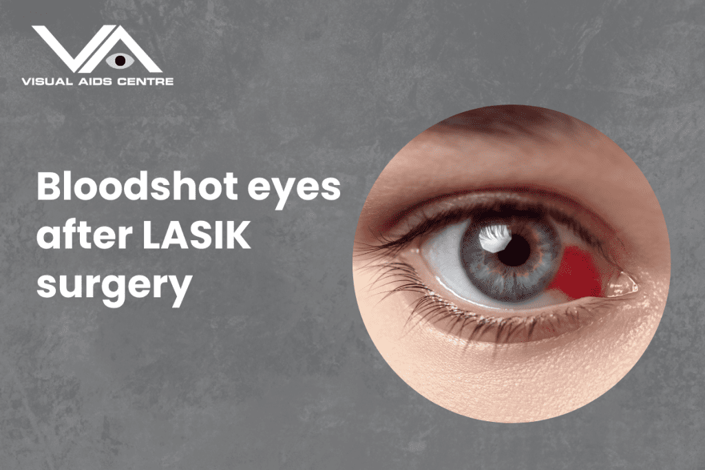
Bloodshot eyes after LASIK surgery are a common and temporary side effect caused by minor trauma to the blood vessels on the surface of the eye during the procedure.
Although they may look alarming, the redness usually resolves on its own within a week or two without affecting your vision.
If you’ve recently had LASIK surgery or are considering it, understanding this phenomenon can give you peace of mind. Here’s a detailed breakdown to help you understand why bloodshot eyes happen, what to expect, and how to manage them effectively.
Why Do Bloodshot Eyes Occur After LASIK Surgery?
The LASIK procedure entails the reshaping of the cornea to rectify vision. During this process, a device called a suction ring is used to stabilise the eye while the corneal flap is created. This step can momentarily increase pressure in the eye, which might cause small blood vessels on the surface to burst. These burst vessels result in tiny red spots or general redness, medically termed subconjunctival haemorrhages.
Contributing Factors:
- Suction Ring Pressure: The suction ring may apply slight force, temporarily increasing blood vessel pressure in the eye.
- Natural Sensitivity: Some people naturally have more fragile blood vessels, making them more prone to redness or spotting.
- Medications: Patients taking blood-thinning medications or supplements before surgery may experience more pronounced redness.
Rest assured, these broken blood vessels do not indicate any complications and will heal naturally over time.
How Long Does the Redness Persist?
The duration of redness varies between individuals but typically resolves within 7 to 14 days post-surgery. Smaller haemorrhages may fade faster, while larger areas of redness can take slightly longer. The good news is that bloodshot eyes rarely require any intervention and should not affect your vision or recovery process.
It’s important to remember that visible redness is a surface-level issue and does not reflect the health of the underlying eye structures, which remain intact after LASIK.
Caring for Bloodshot Eyes Post-LASIK Surgery
While the redness will heal on its own, there are steps you can take to ensure a smooth recovery and avoid worsening the condition. Here’s how:
1. Follow Post-Operative Instructions
Your surgeon will provide instructions tailored to your recovery. These typically include:
- Using prescribed medicated eye drops to prevent dryness and reduce inflammation.
- Avoiding touching or rubbing your eyes, which could worsen redness or impact healing.
- Wearing protective eyewear, especially when sleeping, to prevent accidental contact.
2. Stay Hydrated
Dry eyes can exacerbate discomfort after LASIK. Ensure you’re drinking enough water to maintain adequate hydration, which helps support overall eye health.
3. Use Artificial Tears
Using preservative-free artificial tears can help alleviate any dryness or irritation. These eye drops promote moisture on the eye’s surface, ensuring comfort as the redness subsides.
4. Avoid Eye Strain
Reduce screen time and take regular breaks to prevent digital strain. If you must use devices, ensure proper lighting and practise the 20-20-20 rule—look 20 feet away every 20 minutes for at least 20 seconds.
5. Shield Your Eyes
Protect your eyes from irritants such as dust, smoke, and wind. Sunglasses are a great way to shield your eyes during daytime outings. Preventing further irritation is key to faster healing.
6. Maintain a Healthy Diet
Foods rich in vitamins A and C, along with omega-3 fatty acids, can support tissue healing. Incorporate leafy greens, citrus fruits, and fatty fish like salmon into your diet for optimal eye recovery.
When Should You Be Concerned?
While redness is a normal post-surgical occurrence, some signs may indicate a need for follow-up with your eye surgeon. Contact your doctor immediately if you experience:
- Severe pain: A little discomfort is normal, but sharp or worsening pain may indicate an issue.
- Vision changes: If you notice a decline in vision clarity, blurriness, or spots, seek medical attention.
- Prolonged redness: If the redness persists beyond three weeks, it’s a good idea to have it evaluated.
- Abnormal discharge or swelling: These may suggest an infection or another complication needing timely intervention.
Your doctor is your best resource for assessing any unusual symptoms. Don’t hesitate to reach out for clarity or guidance.
Preventing Bloodshot Eyes After LASIK
Although you cannot entirely prevent redness during LASIK surgery, there are measures you can take before the procedure to minimise its occurrence:
- Disclose any medications, especially blood thinners, to your eye surgeon. They may recommend pausing specific dietary supplements or medications pre-surgery to reduce the risk of haemorrhages.
- Stay calm and relaxed during the procedure. Anxiety can sometimes impact blood vessel fragility.
- Choose an experienced LASIK surgeon with a proven track record for precise, minimally invasive techniques.
Understanding the Healing Process Post-LASIK
Your body is designed to heal efficiently after LASIK, and bloodshot eyes are part of that natural healing process. Over time, the red spots will fade, and your eyes will regain their normal appearance. Though it might feel unsettling to see redness initially, understanding its benign nature can help you stay relaxed.
Throughout your recovery period:
- Focus on overall comfort rather than appearance. Temporary redness doesn’t alter the long-term benefits of LASIK surgery.
- Stay consistent with follow-up appointments. An early check-in with your surgeon ensures everything is progressing as expected.
- Trust the process. LASIK is one of the safest and most effective procedures, with a high rate of patient satisfaction.
The Takeaway on Bloodshot Eyes After LASIK
It’s completely normal to have bloodshot eyes following LASIK surgery. This harmless side effect is the result of burst blood vessels during the procedure, but it poses no threat to your vision or outcome. Most redness resolves within two weeks and requires minimal care beyond following your post-op instructions.
Every patient heals at their own pace, so focus on your eye health and address any concerns directly with your surgeon.
Are you considering LASIK or have questions about recovery? Speak to a trusted eye care specialist to gain clarity and ensure a seamless experience on your vision correction journey.