Three days after LASIK, your vision was improving. Then it suddenly seems blurrier than yesterday. You notice halos, light sensitivity, and a vague discomfort you did not have immediately post-surgery. These are the classic early signs of Diffuse Lamellar Keratitis — and if you are reading this having just noticed them, you need to call your surgeon today, not wait for your next scheduled follow-up.
DLK (first described by Smith and Maloney, 1998) occurs in 0.5–4.3% of LASIK procedures (PMC 2021). Most are Stage 1 and resolve with corticosteroids — the danger is silent progression to Stage 3, where surgical intervention becomes necessary.
Key Takeaways
- DLK occurs in approximately 0.5–4.3% of LASIK cases — most are Stage 1 (mild) and resolve completely with intensive corticosteroid drops.
- Severe DLK (Stages 3 and 4) is rare — approximately 0.016% in large clinical series (published data, PubMed).
- 98.9% of DLK cases respond to increased corticosteroid dosing (5-year study, PMC 2021). Only Stage 3 and above typically require surgical irrigation.
- DLK is not an infection — it is a sterile inflammatory reaction. Antibiotics are not the primary treatment; corticosteroids are.
- Symptoms typically appear within the first 1–5 days post-LASIK — though late-onset DLK triggered by eye infection or injury years later has been reported.
- Delayed treatment of Stage 2 allows rapid progression to Stage 3 or 4 — contact your surgeon the same day symptoms appear.
What Is DLK and Why Does It Happen?
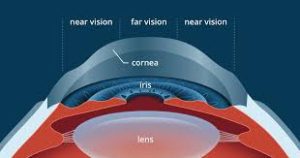
During LASIK, a corneal flap is created and repositioned without sutures. The potential space between the flap and stromal bed is microscopic — but it exists. DLK happens when inflammatory cells migrate into this interface, causing a whitish granular infiltrate visible on slit-lamp examination. It is a sterile immune response — not an infection — triggered by debris, endotoxins, meibomian secretions, or exogenous contaminants. In some cases, conjunctivitis years later can reactivate it. Our guide on LASIK and keratitis risk covers the inflammatory mechanism in detail.
Symptoms to Watch For
Symptoms typically emerge Day 1–5. The earliest sign is reversed improvement — vision that was clearing suddenly blurs again.
| Symptom | Why It Happens | Typical Onset |
|---|---|---|
| Blurry or hazy vision | Inflammatory cell accumulation at the flap interface scatters incoming light | Days 1–5 post-LASIK |
| Light sensitivity (photophobia) | Inflamed corneal tissue increases sensitivity to light stimulus | Days 1–5 |
| Eye discomfort or pain | Inflammatory mediators activate corneal pain receptors | Days 2–5 |
| Eye redness | Vascular dilation from the inflammatory response | Days 2–7 |
| Excessive tearing | Ocular surface irritation triggers lacrimal reflex | Days 1–5 |
None of these symptoms in isolation confirms DLK. What is clinically significant is a reversal of improvement or symptoms worsening after Day 2.
The 4 Stages of DLK — and What Each Means
Stage determines both treatment urgency and method — and progression between stages can happen within 24 hours without treatment.
| Stage | Inflammation Location | Vision Impact | Standard Treatment |
|---|---|---|---|
| Stage 1 | Peripheral only — cells stay outside the central optical zone | Minimal to none | Intensive topical corticosteroids (hourly dosing) |
| Stage 2 | Inflammation extends toward the centre of the cornea | Mild blur, halos | High-dose topical steroids; possible oral corticosteroids; urgent review |
| Stage 3 | Dense central inflammation involving the visual axis | Significant vision reduction | Flap lifting + irrigation of interface + intensive steroids |
| Stage 4 | Severe corneal damage — stromal thinning and scarring | Serious or permanent vision loss | Urgent surgical intervention; outcome dependent on speed of treatment |
Per the PMC 2021 five-year study (14,348 eyes): 76 cases reached Stage 2, 25 Stage 3, and only 3 Stage 4. The critical transition is Stage 1 to Stage 2 — which can occur within 24 hours without treatment.
Causes and Risk Factors
DLK is triggered by exogenous factors (contamination, debris) or endogenous immune responses — both affect the patient’s pre-operative preparation.
- Surgical debris: Particles on blades, endotoxins or detergent residue on instruments — the most common exogenous trigger
- Meibomian gland secretions: Lipid material from eyelid margins entering the flap interface during surgery
- Pre-existing eyelid disease: Blepharitis and MGD significantly increase risk — treat before LASIK
- Dry eye syndrome: Pre-existing dry eye compromises the tear film and increases inflammatory activity at the ocular surface
- Post-operative triggers: Eye infections or trauma months or years after LASIK can reactivate interface inflammation
Treatment by Stage
Per PMC 2021: 98.9% of DLK cases respond to increased corticosteroid dosing. Surgical flap lifting is required only for Stage 3 and above.
- Stage 1: Intensive topical corticosteroid drops — typically hourly dosing for 24-48 hours, then tapering based on clinical response
- Stage 2: Escalated steroid dosing, possible oral corticosteroids, daily clinical review. The prescribed post-LASIK prednisolone drop regimen is intensified significantly at this stage
- Stage 3: Surgical flap lifting, thorough irrigation of the interface to remove inflammatory cells, followed by high-dose topical steroids. Recovery monitored closely post-irrigation
- Stage 4: Urgent intervention — outcome depends entirely on speed of treatment. Some corneal damage at this stage may not be reversible. Long-term vision rehabilitation may be required
When to Seek Immediate Help
Contact your surgeon the same day — do not wait for a scheduled appointment — if you notice any of the following:
- Vision that was improving blurs again after Day 2
- Light sensitivity or pain worsens after initial improvement
- Symptoms worsen despite already being on corticosteroid drops
- Redness increases after the first 48 hours
- Any sudden marked vision drop at any post-LASIK stage
Stage 1 can become Stage 2 in 24 hours; Stage 2 to Stage 3 in another 24 hours. Speed of intervention determines whether vision is preserved. Our LASIK recovery tips guide helps you distinguish normal healing from warning signs.
Prevention Checklist — Before and After LASIK
Modern higher-frequency femtosecond lasers have reduced DLK incidence vs earlier microkeratome techniques (PMC data) — but pre-operative preparation and post-surgery compliance remain equally critical.
| When | Preventive Step | Why It Matters |
|---|---|---|
| 4–6 weeks before LASIK | Treat blepharitis and MGD (meibomian gland dysfunction) | Lipid debris from eyelid margins is a primary flap interface irritant |
| 4–6 weeks before | Stabilise dry eye with lubricating drops | Compromised tear film elevates inflammatory cell activity at the ocular surface |
| Day of surgery | Confirm single-use instruments and powder-free gloves are used | Endotoxin contamination on reusable tools is the primary cause of epidemic DLK outbreaks |
| Post-surgery (all stages) | Use corticosteroid and antibiotic drops exactly as prescribed — never self-taper | Adequate corticosteroid prophylaxis is the primary prevention against post-LASIK interface inflammation |
| Post-surgery | Attend Day 1 and Day 5 follow-ups without exception | Both DLK and its progression to Stage 2 are typically caught at these scheduled examinations |
| Post-surgery (always) | Never rub or press on your eyes | Mechanical pressure physically disrupts the flap interface and can introduce debris from lid margins |
Conclusion
DLK is manageable — 98.9% of cases respond to corticosteroids when caught at Stage 1. What is not manageable is the consequence of delay: Stage 1 costs a visit and a dose change; Stage 4 costs vision. Know the symptoms, act the same day you notice them, and attend every follow-up without exception.
Had LASIK and concerned about a symptom? Book a same-day review at Visual Aids Centre.
Frequently Asked Questions
How common is DLK after LASIK?
Published data: DLK occurs in 0.5–4.3% of LASIK procedures. Most are Stage 1 and resolve with intensive corticosteroid drops. Severe DLK (Stages 3–4) occurs in approximately 0.016% of cases. The “1 in 5,000” figure sometimes cited is a significant underestimate.
What does DLK look like on examination?
Under slit-lamp, DLK appears as a granular white infiltrate at the flap interface — sometimes called “sands of the Sahara.” Unlike infection, there is no discharge and the distribution is diffuse and symmetric rather than focal.
Is DLK an infection?
No — DLK is a sterile inflammatory reaction, not an infection. The primary treatment is corticosteroids to reduce inflammation, not antibiotics. Antibiotics are only added when secondary infection is specifically suspected alongside DLK.
Can DLK cause permanent vision loss?
Stage 1 and 2 — caught and treated promptly — rarely cause lasting vision impact. Stage 3 and 4 can cause corneal scarring and permanent reduction. The single most important factor is how quickly Stage 3 is identified and surgically irrigated.
Can DLK recur after LASIK?
Yes — late-onset DLK has been documented decades post-LASIK, triggered by conjunctivitis or eye injury causing inflammatory cells to migrate to the existing flap interface. Patients should always mention their LASIK history to any future treating ophthalmologist.
What is the treatment for Stage 3 DLK?
Stage 3 requires surgical flap lifting and thorough irrigation to physically remove the inflammatory cell accumulation, followed by intensive topical corticosteroids. This is performed by the treating surgeon and is typically done at a slit-lamp or procedure room — not a full operating theatre.
👁️ MEDICALLY REVIEWED BY
Padmashree Dr. Vipin Buckshey
BS Ophthalmology | AIIMS Graduate, 1977 | Padma Shri Honouree | Refractive Surgery Complications Specialist, Visual Aids Centre
DLK is the post-LASIK complication that most clearly demonstrates why follow-up is not optional. Across more than 250,000 procedures at Visual Aids Centre, Dr. Vipin Buckshey’s consistent observation is that every Stage 3 case that required irrigation was a Stage 1 case not followed up promptly. This article’s data — incidence, staging, and treatment response — is drawn from the PMC 2021 five-year study and the Smith and Maloney 1998 foundational description, cross-referenced with Visual Aids Centre’s clinical experience. An AIIMS alumnus, Padma Shri honouree, and former President of the Indian Optometric Association. Read more at our story.