Yes — the corneal flap is lifted during LASIK surgery. Created using a femtosecond laser (100–120 µm thick) or microkeratome blade (130–160 µm), it is lifted to expose the stromal bed for excimer laser reshaping, then repositioned without sutures. The flap adheres securely but never fuses back to original biomechanical strength — and the implications of this matter for long-term eye care. Here is everything you need to know.
Key Takeaways
- LASIK creates a hinged corneal flap — lifted to expose the stroma for reshaping, repositioned without stitches, and held by natural corneal pressure within hours.
- Two creation methods: femtosecond laser (100–120 µm, more precise) and microkeratome blade (130–160 µm). Femtosecond is standard at most modern LASIK centres.
- The flap adheres but does not fuse permanently — the interface never returns to original stromal biomechanical strength. This is why the flap can be re-lifted years later for enhancement procedures.
- Flap displacement occurs in approximately 0.1–0.5% of cases — most common in the first 24–48 hours. Eye rubbing is the primary avoidable cause.
- Other flap complications: DLK (0.4–2%), epithelial ingrowth (0.2–3.9% primary LASIK), and flap striae (macro-striae ~0.1–0.5%).
- Flapless procedures (SMILE Pro, Trans-PRK, ICL) eliminate flap-specific complications entirely — important for active lifestyle, contact sports, or borderline corneal thickness.
What Is the LASIK Corneal Flap — and Why Is It Lifted?
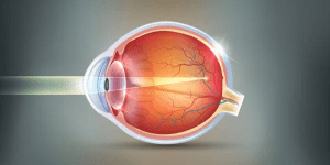
The corneal flap is a thin, hinged disc — typically 8.5–9.5 mm in diameter — cut through the epithelium into the anterior stroma. The hinge (most commonly nasal with femtosecond, or superior with some microkeratome systems) keeps the flap attached during lifting and repositioning.
Once lifted, the flap exposes the stroma for excimer reshaping. After ablation it is repositioned without sutures — IOP and epithelial surface tension hold it in place within minutes, enabling LASIK’s 24–48 hour recovery.
How the LASIK Flap Is Created — Femtosecond Laser vs Microkeratome
Our guide on bladeless vs blade LASIK covers the detailed outcome data for both creation methods.
| Feature | Femtosecond Laser (Bladeless) | Microkeratome (Blade) |
|---|---|---|
| Flap thickness | 100–120 µm — thinner; more residual stroma preserved for ablation | 130–160 µm — thicker; slightly less residual stroma post-ablation |
| Flap diameter | Precisely programmable (typically 8.5–9.5 mm) | Determined by blade head size; less customisable |
| Hinge position | Typically nasal (biomechanically preferred) | Superior or nasal depending on blade head |
| Edge architecture | Side-cut angle programmable (vertical, bevelled) — cleaner edge; better flap adhesion | Angled edge from blade geometry — slightly less precise adhesion |
| Corneal hysteresis impact | Less CH reduction — thinner flap removes less biomechanically critical anterior stroma | Greater CH reduction — thicker flap removes more anterior stroma |
| Standard of care | Current standard at most modern centres | Still used; effective but older technology |
What Happens After the Flap Is Lifted?
With the flap on its hinge, the excimer laser ablates the stromal bed — approximately 12–14 µm of tissue per dioptre corrected (a -5.00D correction removes ~60–70 µm).
After ablation, the surgeon irrigates and repositions the flap — no sutures. IOP holds it in place; epithelial bridging begins within 2–4 hours. By 24 hours most patients can resume normal activities.
The flap never fuses to original stromal strength — the interface reaches ~28% of native strength by 3 months, remaining lower indefinitely. This is why the flap can be re-lifted for LASIK enhancement years or decades later.
LASIK Flap Complications — Types, Incidence and Management
Flap-related complications are uncommon with modern technique — but understanding their incidence and management matters for informed consent. Our guide on LASIK flap dislocation covers how to recognise and respond to displacement specifically.
| Complication | Published Incidence | Presentation | Management |
|---|---|---|---|
| Flap displacement | ~0.1–0.5% of cases; most common first 24–48 hours | Sudden blurring, distortion, or discomfort; patient may notice flap has shifted | Urgent review — flap irrigation, repositioning, hydration. Early presentation allows full visual recovery. |
| Diffuse Lamellar Keratitis (DLK) | 0.4–2% of LASIK cases; mild cases Grade 1 are most common and self-limiting | Interface haze/granular appearance (“sands of the Sahara”) on slit lamp; reduced contrast sensitivity; may progress without treatment | Grade 1–2: intensive topical steroids. Grade 3–4: flap lift, irrigation, and systemic steroids. Early detection critical — DLK progresses rapidly without treatment. |
| Epithelial ingrowth | 0.2–3.9% primary LASIK; up to 18–19% re-treatment LASIK | Grey-white opacity at flap edge; blurring or irregular astigmatism if approaching visual axis | Grade 1: observe. Grades 2–3: flap lift + debridement + adjuvant therapies (MMC, fibrin glue) |
| Flap striae (wrinkles) | Micro-striae: common, usually insignificant. Macro-striae: ~0.1–0.5% | Micro-striae: slit lamp only, no functional impact. Macro-striae: irregular astigmatism, visual disturbance | Micro-striae: observe. Macro-striae: flap lift, stretch, re-adhesion. Earlier intervention = better result. |
LASIK (Flap) vs Flapless Procedures — Which Is Right for You?
For patients where the flap is a concern — contact sports, thin corneas, active lifestyle, or dry eye — flapless alternatives deserve serious consideration:
| Factor | LASIK (Flap) | SMILE Pro (Flapless) | Trans-PRK (Surface) | ICL (No ablation) |
|---|---|---|---|---|
| Flap created? | ✅ Yes — lifted and repositioned | ❌ No flap — small 2–4 mm incision | ❌ No flap — surface ablation | ❌ No corneal tissue removed |
| Dry eye risk | Moderate — flap severs corneal nerves; regeneration 3–6 months | Lower — incision preserves more corneal nerve integrity | Moderate — surface ablation; slower nerve regeneration | Minimal — no corneal tissue altered |
| Contact sports / eye rubbing | Higher risk — flap can be displaced by trauma; long-term eye rubbing concern | Lower — small incision heals fully; no large flap to displace | No flap risk — surface heals completely; still avoid rubbing | Lowest — no corneal modification; eye rubbing not a structural risk |
| Best for | Most patients with adequate corneal thickness prioritising fast recovery | Active lifestyle, contact sports, dry eye, borderline biomechanics — same correction range as LASIK | Thin corneas, military/police candidates (no flap required) — longer recovery acceptable | High myopia (>−8D), thin corneas, unstable prescriptions — reversible procedure |
Post-LASIK Flap Protection — What to Do and Avoid
The flap is most vulnerable in the first week. For questions about specific post-op events — like accidentally touching the eye — our guide on poking your eye after LASIK covers when to be concerned and when not to worry.
| Timeframe | ✅ Do | ❌ Avoid | Why It Matters for Flap |
|---|---|---|---|
| Day 1 (first 24 hours) | Wear prescribed protective shields; use lubricating drops; rest eyes | No rubbing, pressing, or touching eyes; no bending head below waist; no strenuous activity; no hot showers near eyes | Flap least adherent in first 24 hours — minimal force can displace it at this stage |
| Days 2–7 | Continue antibiotic + steroid drops; shields while sleeping; attend follow-up; resume light activity | Eye rubbing; swimming; contact sports; dusty/smoky environments; eye makeup | Epithelial bridging underway but incomplete — pressure or contamination can still displace the flap or cause infection |
| Weeks 2–4 | Switch to preservative-free lubricating drops; regular follow-up; moderate exercise | Swimming without goggles; contact sports without eye protection; eye rubbing | Flap edges healing but interface not yet stable — impact or contaminated water at flap margin is a displacement and infection risk |
| Month 1 onwards | Resume all activities including swimming (goggles initially); UV-protective sunglasses outdoors; 1-month review | Eye rubbing indefinitely — the flap never reaches original biomechanical strength; habitual rubbing remains a long-term risk | Flap is stable but not permanently fused — trauma sufficient to displace it remains possible years post-surgery, though risk decreases substantially after month 1 |
Conclusion
The LASIK flap (100–120 µm femtosecond; 130–160 µm microkeratome) is lifted to expose the stromal bed, repositioned without sutures, and heals securely — but never fuses to original biomechanical strength. Principal risks (displacement ~0.1–0.5%; DLK 0.4–2%; ingrowth 0.2–3.9%) are manageable with early detection. For patients where flap risk is a concern, SMILE Pro and Trans-PRK eliminate it entirely.
Considering LASIK and want to understand whether a flap-based or flapless procedure is right for your cornea? Book a consultation at Visual Aids Centre — full pre-operative workup including Pentacam, corneal thickness, and procedure recommendation included.
Frequently Asked Questions
Is the flap lifted during LASIK surgery?
Yes — the flap (8.5–9.5 mm diameter; 100–120 µm with femtosecond laser) is lifted to expose the stromal bed for excimer reshaping, then repositioned without sutures. IOP and epithelial bridging hold it in place within hours.
Does the LASIK flap heal permanently?
Securely but not permanently — the interface reaches ~28% of native strength by 3 months and remains lower indefinitely. This enables re-lifting for enhancement procedures years later; it also explains why eye rubbing is a permanent long-term flap risk.
Femtosecond vs microkeratome — what’s the difference for flap creation?
Femtosecond creates a thinner flap (100–120 µm) vs microkeratome (130–160 µm). Thinner = more residual stroma for ablation, less corneal hysteresis reduction, better flap edge architecture. Femtosecond is the current standard of care at most modern centres.
What happens if the LASIK flap is displaced?
Displacement (~0.1–0.5% of LASIK cases; most common in the first 24–48 hours) causes sudden blurring or distortion. Contact your surgeon immediately — prompt irrigation and realignment restores full vision when treated early. Avoiding eye rubbing is the key prevention measure.
What flapless alternatives to LASIK exist?
SMILE Pro (lenticule via 2–4 mm incision), Trans-PRK (surface ablation, no flap), and ICL (implant, no corneal tissue removed) all avoid the flap entirely — eliminating displacement, DLK, ingrowth, and striae risks. Best for active lifestyles, contact sports, or borderline corneal thickness.
👁️ MEDICALLY REVIEWED BY
Padmashree Dr. Vipin Buckshey
BS Ophthalmology | AIIMS Graduate, 1977 | Padma Shri Honouree | LASIK Procedure Specialist, Visual Aids Centre
Femtosecond flap thickness (100–120 µm), microkeratome (130–160 µm), flap diameter (8.5–9.5 mm), displacement (~0.1–0.5%), DLK (0.4–2%), ingrowth (0.2–3.9% primary; up to 18–19% re-treatment), macro-striae (~0.1–0.5%), interface strength (~28% native at 3 months), and ablation rate (~12–14 µm/dioptre) are sourced from published refractive surgery literature including IntraLase femtosecond clinical data and complication management series. The non-permanent fuse position reflects corneal wound healing literature — the interface never achieves original stromal strength, enabling re-lifting for enhancements. Patients at Visual Aids Centre receive full explanation of flap technique, thickness planning, and post-op care during pre-operative consultation. An AIIMS alumnus, Padma Shri honouree, and former President of the Indian Optometric Association. Read more at our story.