If you are preparing for a government, defence, or railway medical examination and considering Contoura Vision surgery to correct your vision, the detectability question is almost certainly the first one on your mind. The detailed answer matters — because it determines which procedure you choose, when you schedule it relative to your exam, and how you navigate the disclosure conversation if one is required.
This guide from Visual Aids Centre gives you a clinically honest answer. Contoura Vision is significantly harder to detect than flap-based LASIK in standard medical examinations — but “harder to detect” is not the same as “undetectable.” Understanding the distinction, what standard examinations can and cannot find, and what advanced equipment can reveal, is what allows you to make this decision with full information rather than assumptions.
Key Takeaways
- Contoura Vision is a flapless surface ablation procedure. It creates no corneal flap and leaves no flap edge — the most visible sign of LASIK under standard slit lamp examination.
- In standard government medical examinations — refraction, fundoscopy, colour vision, and basic slit lamp — Contoura Vision performed 2–3 months prior typically goes undetected.
- Advanced equipment (Pentacam, high-definition topography, corneal tomography) can detect Contoura Vision by identifying the characteristic corneal thinning and curvature changes produced by any ablative procedure.
- For examinations at facilities that use advanced diagnostic equipment — particularly railways, DGCA (aviation), and some higher-tier defence medicals — Contoura Vision may be identifiable with sufficient diagnostic rigour.
- Honest disclosure remains the clinically and professionally correct approach regardless of procedure type or detectability profile.
What Is Contoura Vision?
Contoura Vision is a topography-guided surface ablation laser procedure — a form of advanced PRK (Photorefractive Keratectomy) that uses detailed corneal mapping data to create a highly personalised ablation profile. Unlike standard LASIK, which creates a corneal flap to access the underlying stroma, Contoura Vision removes the epithelial layer and applies the laser correction directly to the corneal surface. No blade is used. No incision is created. No flap is formed.
The procedure corrects myopia, hyperopia, and astigmatism — including higher-order aberrations that standard laser procedures cannot address — by reshaping the corneal surface according to a topographic map unique to each patient’s eye. Our overview of Contoura Vision eye surgery covers the complete procedure, candidacy criteria, and expected outcomes for patients who are new to the concept.
The important distinction for detectability purposes is this: standard LASIK = flap + ablation. Contoura Vision = ablation only, no flap. This architectural difference is the primary driver of the detectability difference between the two procedures.
Why Contoura Vision Is Harder to Detect Than LASIK
Standard flap-based LASIK leaves a permanent, visible diagnostic signature that requires no specialised equipment to identify: the flap edge. Under slit lamp illumination, the boundary between the repositioned corneal flap and surrounding uncut tissue presents as a distinct circumferential ring. Any ophthalmologist performing a routine eye examination sees this finding immediately. LASIK is, in this sense, self-declaring.
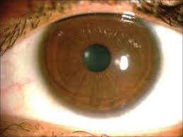
Contoura Vision removes this finding entirely. There is no flap. There is no flap edge. There is no interface between repositioned and uncut tissue. Under standard slit lamp examination at the magnifications and illumination techniques typically used in government medical examinations, a healed Contoura Vision procedure leaves no visible corneal surface abnormality.
What it does leave — permanently — is a measurable change in corneal curvature and thickness. The ablation that corrects your vision removes real corneal tissue. That tissue is gone, the central cornea is thinner, and its curvature profile has changed in a characteristically ablative pattern. These changes are not visible to the naked eye or standard slit lamp — but they are quantifiable by instruments designed to measure them. Our detailed comparison of Contoura Vision versus LASIK covers the full procedural and structural differences that drive this detectability gap.
What Standard Government Medical Examinations Check
The standard ophthalmic battery in most Indian government recruitment medicals — including SSC, railways, state police, army, and many paramilitary services — typically includes:
- Refraction: Measurement of spectacle power. Post-Contoura Vision, this typically reads zero or near-zero — which is what an examiner wants to see, not a sign of surgery.
- Visual acuity testing: Snellen chart assessment at distance and near. Post-Contoura Vision, acuity meets or exceeds the required standard.
- Colour vision assessment: Ishihara plates or equivalent. Not affected by refractive surgery.
- Fundoscopy: Retinal examination. Not affected by Contoura Vision.
- Basic slit lamp examination: Anterior segment assessment at the magnification and technique typically used in general medical examinations. Does not reliably detect flapless surface ablation.
At facilities using only this standard battery, Contoura Vision performed 2–3 months prior to the examination is consistently not identified. The examiner finds an eye that sees clearly without correction — which is precisely what they are looking for. For a detailed understanding of what specific service categories require and how vision standards apply, our guide on laser eye surgery eligibility for the Indian Air Force gives a relevant benchmark of how vision correction history intersects with service requirements.
What Advanced Equipment Can Detect
The corneal changes produced by Contoura Vision — thinning and curvature modification — are detectable by instruments designed to quantify them. Three specific instruments are relevant:
Pentacam / Scheimpflug Tomography
Pentacam generates precise pachymetric maps showing corneal thickness at every point. A Contoura Vision-treated eye shows characteristic central thinning that is diagnostically distinct from natural thin cornea presentations. The Scheimpflug cross-sectional images also reveal the ablation profile geometry in the anterior stroma.
High-Definition Corneal Topography
Topographic maps of Contoura Vision-treated corneas show the characteristic flattening pattern in the central optical zone that follows surface ablation. This pattern is distinguishable from normal corneal variation by an experienced examiner reviewing the map.
Wavefront Aberrometry
The post-ablation wavefront profile — with its characteristic spherical aberration changes — provides supporting evidence of prior laser surgery even when topographic changes are subtle.
Facilities that routinely use these instruments as part of the ophthalmic examination — which includes certain DGCA aviation medicals, railway divisional medical centres, and some higher-tier defence medical boards — can potentially identify Contoura Vision. The probability of detection increases with the sophistication of the diagnostic equipment available.
Detectability by Examination Type
| Examination Type | Standard Equipment | Advanced Equipment | Contoura Detectability |
|---|---|---|---|
| SSC / State Police | Yes | Usually not | Low |
| Army / Paramilitary | Yes | Varies by medical centre | Low to Moderate |
| Railways (divisional) | Yes | Higher chance of HD slit lamp | Moderate |
| DGCA Aviation | Yes | Yes — Pentacam often used | High |
| Navy / Special Forces | Yes | Yes — specialist centres | High |
For candidates targeting services where advanced equipment is used as standard, it is worth understanding what Contoura Vision eligibility specifically means for that service. Our resource on Contoura Vision for government job candidates covers service-specific eligibility and detectability considerations across the most common examination categories.
Timing Contoura Vision Before a Medical Examination
Recovery after Contoura Vision differs from LASIK recovery. Because the epithelium is removed and regenerates — rather than being preserved as a flap — initial vision recovery takes longer. Most patients have functional distance vision within five to seven days, but the corneal surface continues remodelling for several weeks. Full stability of the refractive result and complete surface maturation typically takes two to three months.
For government medical examinations, the timing recommendation is a minimum of two to three months between Contoura Vision surgery and the examination date. This serves two purposes: first, the corneal surface is fully healed, giving the most stable and natural-appearing result. Second, the examination occurs when the refraction is fully settled and the corneal appearance has reached its most stable, least surgically obvious state.
Our detailed resource on Contoura Vision recovery time maps the exact week-by-week progression — confirming when vision stabilises, when the surface is fully healed, and what the examination milestones look for at each stage.
Clinical Advantages of Contoura Vision for Government Job Candidates
Beyond detectability, Contoura Vision offers genuine clinical advantages that make it a strong choice for the government job candidate population:
- Flapless architecture: No corneal flap means no risk of flap displacement from physical activity, training, or trauma — permanently. Relevant for candidates who will undertake vigorous physical training as part of their role.
- Suitable for thin corneas: Candidates whose corneas are insufficiently thick for LASIK may still qualify for Contoura Vision, which can be performed on thinner corneal profiles than flap-based procedures.
- Higher-order aberration correction: Contoura’s topography-guided ablation corrects not just the primary prescription but the corneal irregularities that affect contrast sensitivity and night vision — both relevant to operational roles.
- Better night vision outcomes: The topography-guided approach consistently produces better low-light visual quality than standard LASIK for comparable prescriptions.
For candidates whose corneal profile specifically makes them suitable for Contoura Vision over standard LASIK — including those with thin corneas or irregular topography — our resource on Contoura Vision for thin corneas covers the specific thickness parameters and what the procedure can achieve for patients in this category.
Post-Operative Care and Recovery
Contoura Vision’s recovery period is longer and initially more uncomfortable than LASIK — the trade-off for the flapless architecture. Key post-operative milestones:
- Days 1–5: Vision is blurred as the epithelium regenerates. Protective lenses are worn during this phase. Significant light sensitivity is common.
- Week 1–2: Functional vision returns for most daily tasks. Screen use is permitted but reduced.
- Month 1: Corneal surface is largely healed. Residual fluctuation in vision quality is normal as the stroma remodels.
- Month 2–3: Full visual stability is typically achieved. This is the minimum window before government medical examinations.
The outcomes data for Contoura Vision — including what percentage of patients achieve 6/6 vision or better and how results compare to standard LASIK — is covered in our resource on Contoura Vision success rate.
On Disclosure — The Only Advice That Matters Long-Term
This section needs to be stated clearly, because the detectability discussion in this article could otherwise be misread as a guide to concealment. It is not. Visual Aids Centre’s position is unambiguous: honest disclosure of surgical history during government and defence medical examinations is always the correct approach.
Contoura Vision’s lower detectability profile is clinically relevant for candidates appearing for services that do not specifically ask about vision correction surgery — where the question of disclosure simply does not arise. For services that specifically ask whether any refractive surgical procedure has been performed, or that require comprehensive ophthalmological history, concealment is misrepresentation of medical history regardless of how detectable the procedure is. The consequences of discovered concealment — which can occur at any stage of a career, including well after selection — are more severe than any disclosed surgical history.
The correct use of the information in this article is to choose the procedure that best fits your clinical profile, time it appropriately before your examination, and ensure your vision meets the required standard at the time of examination. Book a consultation at Visual Aids Centre to understand which procedure gives you the best clinical outcome for your specific vision correction needs and examination context.
Conclusion
Contoura Vision is genuinely harder to detect than standard LASIK in most government medical examinations — primarily because it creates no corneal flap and leaves no flap edge. At facilities using standard examination protocols (refraction, fundoscopy, basic slit lamp), Contoura Vision performed 2–3 months prior is typically not identified. At facilities using advanced diagnostic equipment — Pentacam, high-definition topography, wavefront aberrometry — the ablation-induced corneal changes can be identified by an experienced examiner.
Choose the procedure that gives you the best clinical outcome for your vision and examination context. Time it appropriately. Disclose honestly when asked. And arrive at your medical examination with vision that meets the required standard on its own merits.
Frequently Asked Questions (FAQs)
Is Contoura Vision detectable in government medical exams?
At facilities using standard examination protocols (refraction, colour vision, basic slit lamp), Contoura Vision is typically not detected, particularly when performed 2–3 months before the examination. Advanced diagnostic equipment (Pentacam, HD topography) can detect the characteristic corneal changes.
Why is Contoura Vision harder to detect than LASIK?
LASIK creates a corneal flap that leaves a permanently visible flap edge — identifiable under basic slit lamp examination by any ophthalmologist. Contoura Vision is flapless; it leaves no flap edge. The only detectable changes are corneal thinning and curvature modification, which require specialised instruments to quantify.
How long before a government medical should I have Contoura Vision?
A minimum of 2–3 months. This allows the corneal surface to heal completely, the refraction to stabilise fully, and the corneal appearance to reach its most stable, least surgically obvious state.
Can Contoura Vision be detected by Pentacam?
Yes. Pentacam generates precise corneal thickness maps that reveal the characteristic thinning pattern from surface ablation. At facilities using Pentacam as standard — certain railway, aviation, and specialised defence medicals — Contoura Vision can be identified by experienced examiners.
Should I disclose Contoura Vision in government medical exams?
If the examination specifically asks whether any refractive procedure has been performed, honest disclosure is always the correct response. Concealment is misrepresentation of medical history — the consequences of discovery are more severe than those of honest disclosure. If the examination does not ask, the question does not arise.
Is Contoura Vision better than LASIK for candidates appearing for government medicals?
For examinations using only standard equipment, Contoura Vision has a lower detectability profile than LASIK. It also offers genuine clinical advantages — flapless architecture, thin cornea suitability, and better night vision outcomes. For high-scrutiny examinations using advanced equipment, detectability differences narrow considerably.
👁️ MEDICALLY REVIEWED BY
Padmashree Dr. Vipin Buckshey
BS Ophthalmology | AIIMS Graduate, 1977 | Padma Shri Honouree | Government Examination Medical Navigation Specialist, Visual Aids Centre
The intersection of refractive surgery choice and government medical examination requirements is one of the most practically consequential consultations Visual Aids Centre conducts. Dr. Vipin Buckshey has guided candidates across virtually every major Indian government and defence recruitment medical category — understanding what each examining facility’s diagnostic capability actually is, how different procedures interact with those capabilities, and how candidates can make genuinely informed decisions rather than decisions based on assumptions. The clinical accuracy in this article — particularly regarding what different examination facilities can and cannot detect — reflects that direct experience at Visual Aids Centre rather than general estimates. An AIIMS alumnus, Padma Shri honouree, and former President of the Indian Optometric Association. Read more about our approach at our story.